Introduction to normalization
The problem
What are we trying to match?
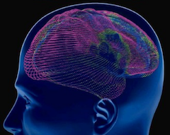
Anatomical variation
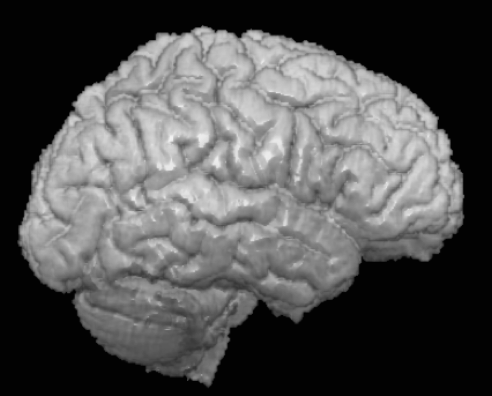
Even identical twins have different sulcal anatomy (Klein & Tourville, 2012). There are renderings of the left hemisphere of two identical twins.

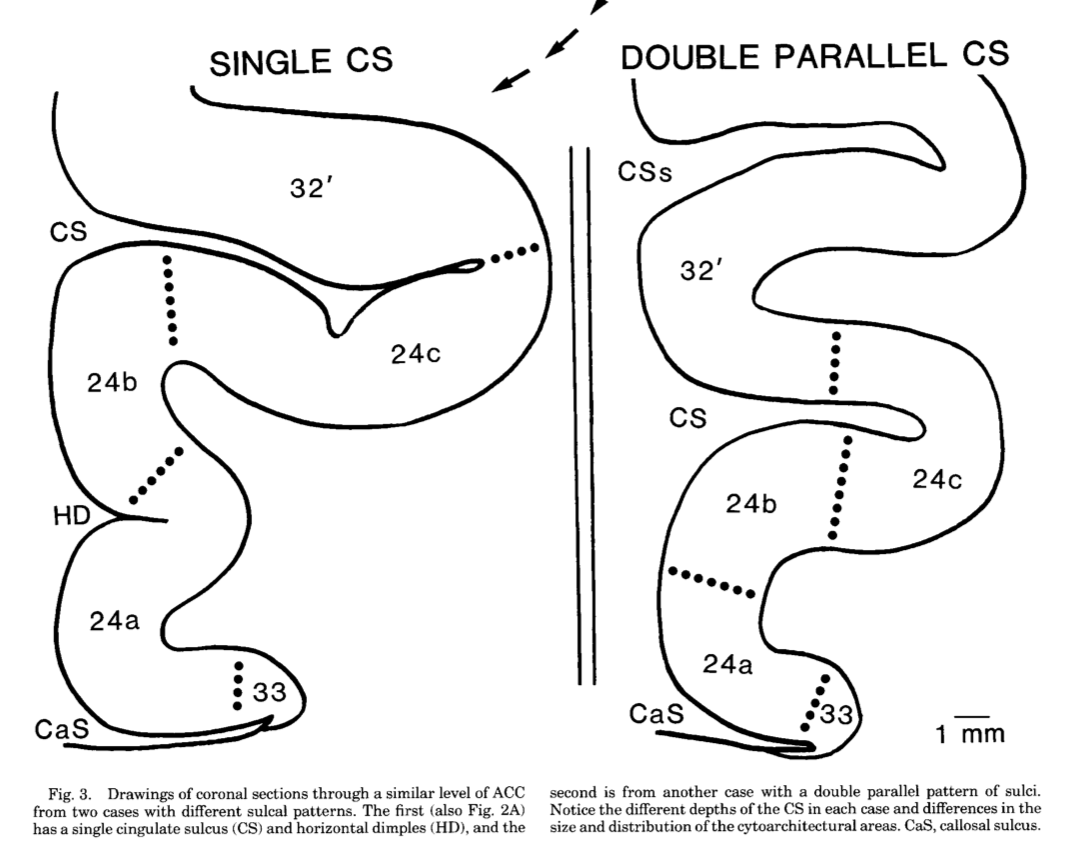
Anterior cingulate
See (Vogt, Nimchinsky, Vogt, & Hof, 1995)
Most brain hemispheres (65%) have a single cingulate gyrus delimited by a cingulate sulcus above it:
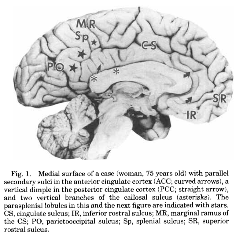
The rest have two cingulate sulci, with an extra superior cingulate sulcus and superior cingulate gyrus:
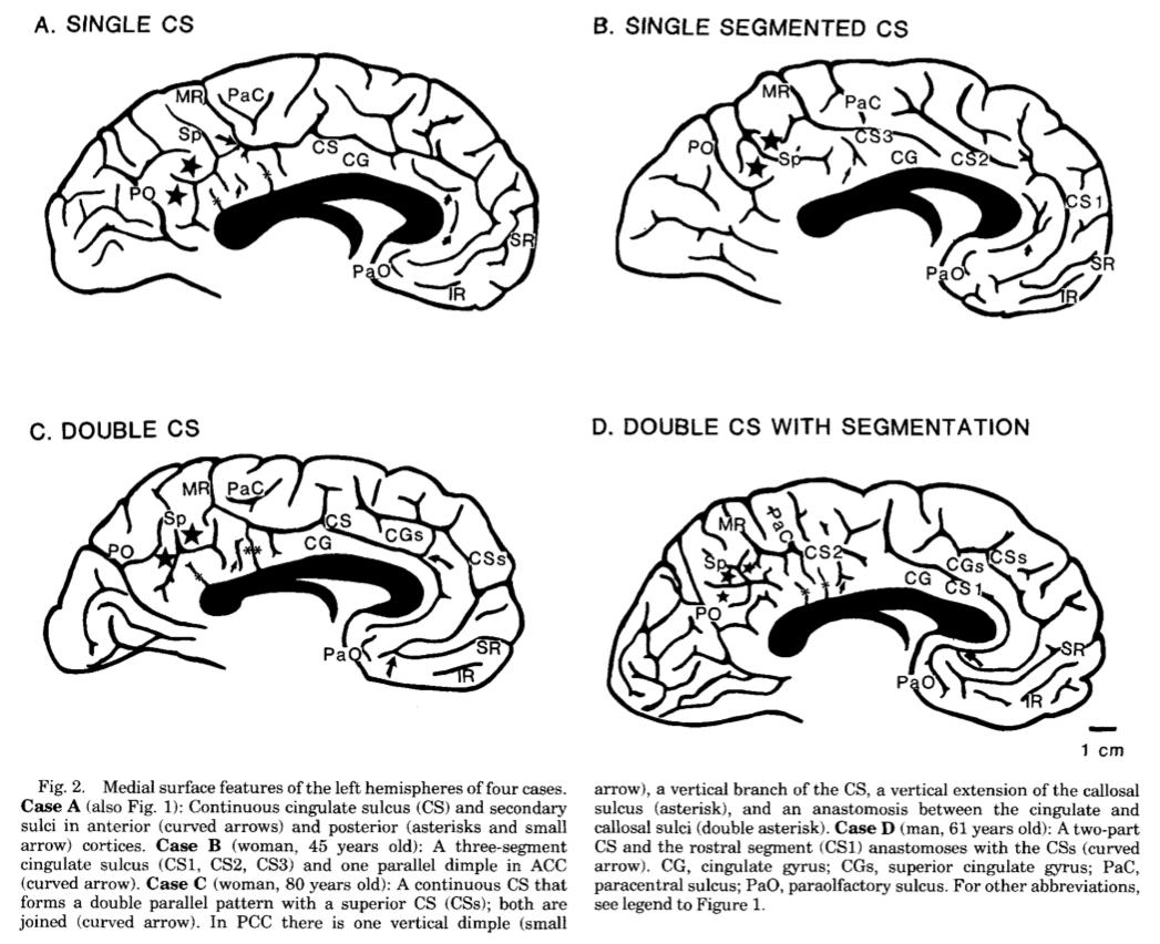
The cytoarchitectonic regions appear to correspond to the sulcal anatomy:
Matching to a not-individual template
Our problem is often worse than that. We are often matching to a template brain that is the average of lots of brains, and has therefore lost much of its anatomical detail.
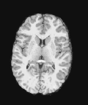
Individual brain
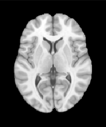
Template brain
Sulcal matching
Material from the following slides kindly provided by Michael Waskom.
Freesurfer software tries to match brains using their sulci and gyri.
The first step is detecting, and then inflating the cortical surface:
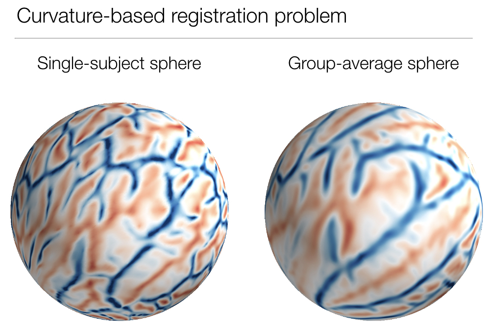
Freesurfer keeps the curvature information with the surface positions, after inflating to a sphere. In the graphics here, convex areas are in blue. As you see, these are the tops of gyri. Convex areas are in red, and these are characteristic of the bottom of sulci.
It then matches the individual curvature pattern to a template of averaged curvature patterns.
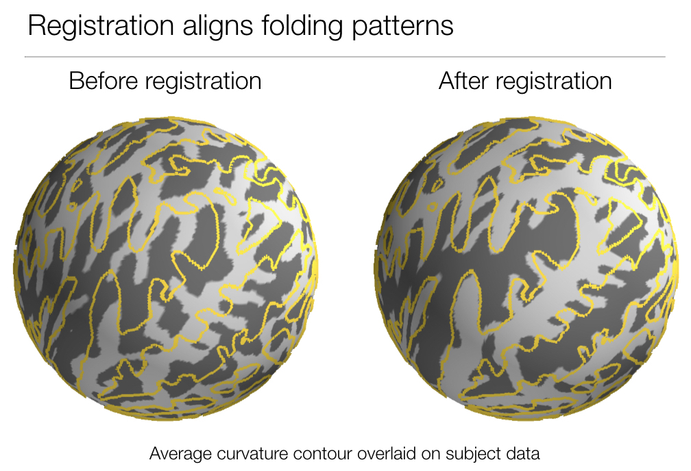
This is an animation of the distortion process resulting from matching to the template:
The registration aligns sulcal patterns:
Here are some graphics from (Fischl, Sereno, Tootell, & Dale, 1999), showing the difference between standard greyscale matching of the image volumes (“Talairach” matching) and sulcal matching (with Freesurfer).
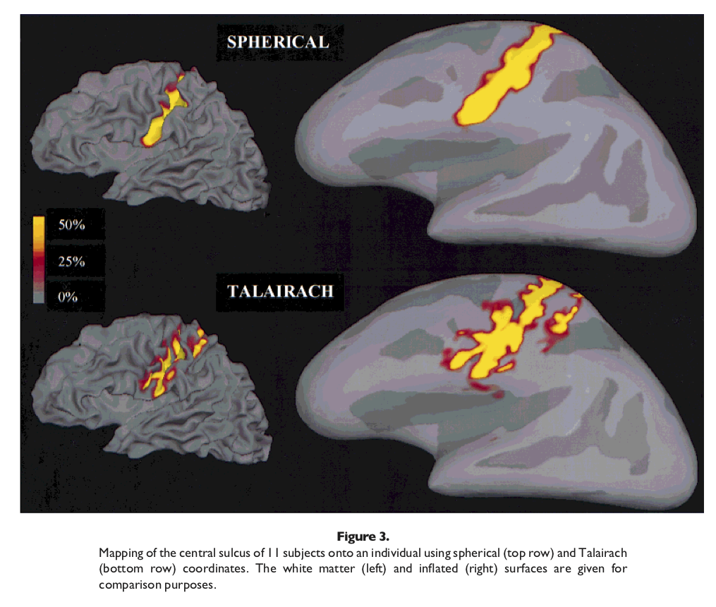
Freesurfer / Talairach comparison, motor cortex
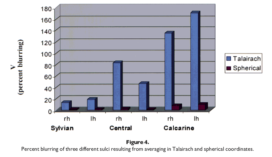
Sulcal blurring with motor cortex
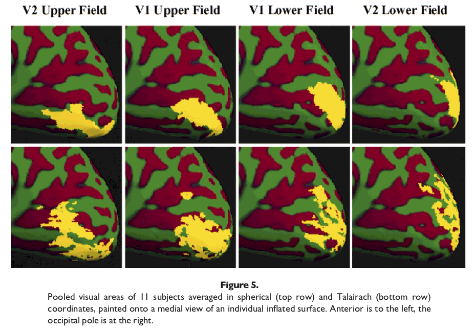
Freesurfer / Talairach, visual cortex
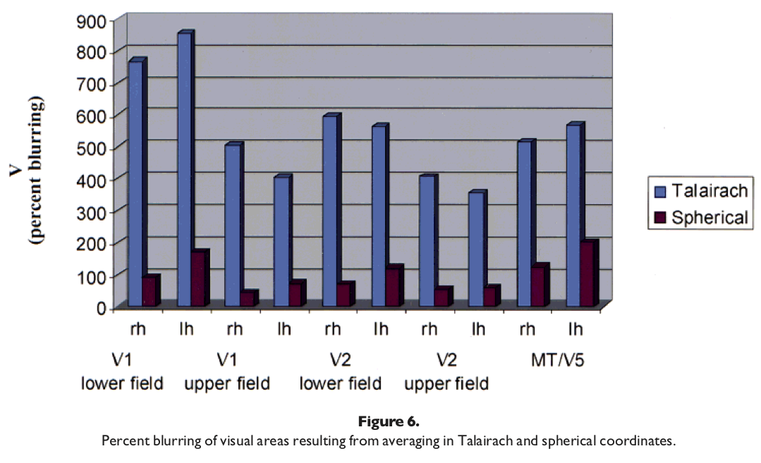
Sulcal blurring with visual cortex
See also
References
- Klein, A., & Tourville, J. (2012). 101 labeled brain images and a consistent human cortical labeling protocol. Frontiers in Neuroscience, 6, 171.
- Vogt, B. A., Nimchinsky, E. A., Vogt, L. J., & Hof, P. R. (1995). Human cingulate cortex: surface features, flat maps, and cytoarchitecture. Journal of Comparative Neurology, 359(3), 490–506.
- Fischl, B., Sereno, M. I., Tootell, R. B. H., & Dale, A. M. (1999). High-resolution intersubject averaging and a coordinate system for the cortical surface. Human Brain Mapping, 8(4), 272–284.